
Neuron Model Manufacturer,Supplier and Exporter in India
Product Code : JA-AM-205
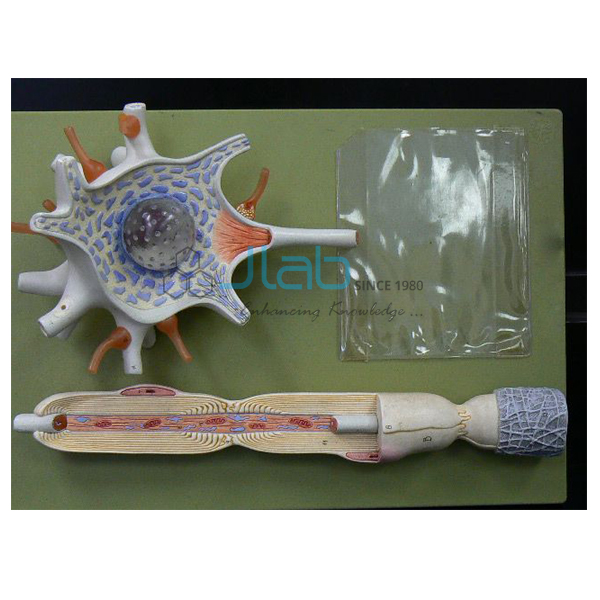
The Neuron Model from Jaincolab consists of 2 parts: the neuronal body and the nerve fiber, giving students a detailed view of the internal and peripheral structures of a nerve cell across 15 labeled positions.
This neuron anatomical model is molded from advanced PVC and painted with imported paint. The neuronal body section shows the perikaryon cut open to expose the nucleus of the cell, the neurite cone, dendrites and flexible synaptic terminals, along with the axon hillock, dendrite arbour and neurofilament.
The nerve fiber section shows the peripheral nerve with its sheaths, including the Schwann cell with its nucleus, the Schwann sheath, the axon and mitochondria. As a trusted anatomical models supplier in India, Jaincolab manufactures this neuron model to support neuroanatomy teaching in schools, colleges and medical training institutes across India and internationally.
Product Specifications
| Parameter | Details |
|---|---|
| Parts | 2 parts: neuronal body and nerve fiber |
| Material | Advanced PVC, painted with imported paint |
| Labeled Positions | 15 positions displayed |
| Neuronal Body Shows | Perikaryon (cut open), nucleus, neurite cone, dendrites, synaptic terminals, axon hillock, dendrite arbour, neurofilament |
| Nerve Fiber Shows | Peripheral nerve with sheaths: Schwann cell and nucleus, Schwann sheath, axon, mitochondria |
| Product Code | JA-AM-205 |
Key Features
- 2-part model: neuronal body and nerve fiber sections
- Shows axon hillock, dendrite arbour and neurofilament in detail
- Perikaryon cut open to expose nucleus, neurite cone, dendrites and synaptic terminals
- Peripheral nerve section shows Schwann cell, Schwann sheath, axon and mitochondria
- 15 labeled positions for precise structural identification
- Made of advanced PVC with imported paint for durability and finish
Why Choose Jaincolab
Jaincolab is a well-established anatomical models supplier in India, serving schools, colleges and medical training institutes across India and internationally. This neuron model complements Jaincolab's Brain with Arteries on Head and Cranial Nerve Skull for complete neuroanatomy teaching, within the wider Anatomical Models Equipments range. Jaincolab is ISO 9001:2015 and ISO 14001:2015 certified.
Applications
| Application Area | Use |
|---|---|
| Neuroanatomy Teaching | Studying the detailed structure of a neuron cell body and its nerve fiber |
| Medical & Nursing Institutes | Reference model for nerve cell and peripheral nerve anatomy |
| Biology Laboratories | Detailed teaching aid for nervous system lessons |
| Science Exhibitions | Enlarged, labeled model for interactive classroom demonstration |
Frequently Asked Questions
Q1. What parts does the Neuron Model consist of?
A. It consists of 2 parts: the neuronal body and the nerve fiber.
Q2. What structures are shown on the neuronal body section?
A. It shows the perikaryon cut open to expose the nucleus, neurite cone, dendrites, synaptic terminals, axon hillock, dendrite arbour and neurofilament.
Q3. What does the nerve fiber section show?
A. It shows the peripheral nerve with its sheaths, including the Schwann cell and nucleus, Schwann sheath, axon and mitochondria.
Q4. What material is the model made of?
A. It is made of advanced PVC and painted with imported paint.
Q5. How many labeled positions does this model display?
A. The model displays 15 labeled positions.
Request a Quote
To request pricing or place an order for the Neuron Model, please contact our team with your requirement.


